Nanolive introduces world’s first holo-tomographic fluorescent microscope
One year after launching its unique 3D Cell Explorer (the first microscope able to look inside living cells without harming them, in 3D and real time), the EPFL spin-off reaches a new fundamental milestone for cell biology and microscopy and releases its latest pioneering product: the 3D Cell Explorer-fluo.
The holo-tomographic technology proper of the 3D Cell Explorer allows to measure the quantitative refractive index (RI) of the cells’ organelles with nanometric precision and instantly. The technology was published in Nature Photonics in 2013, and the 3D Cell Explorer was already awarded with multiple prestigious awards (e.g. Top 10 Innovation of 2015 from The Scientist or R&D100).
THE MOST POWERFUL SOLUTION TO STUDY LIVING CELLS: COMBINING CELL STRUCTURE WITH SPECIFIC FLUORESCENT MARKERS
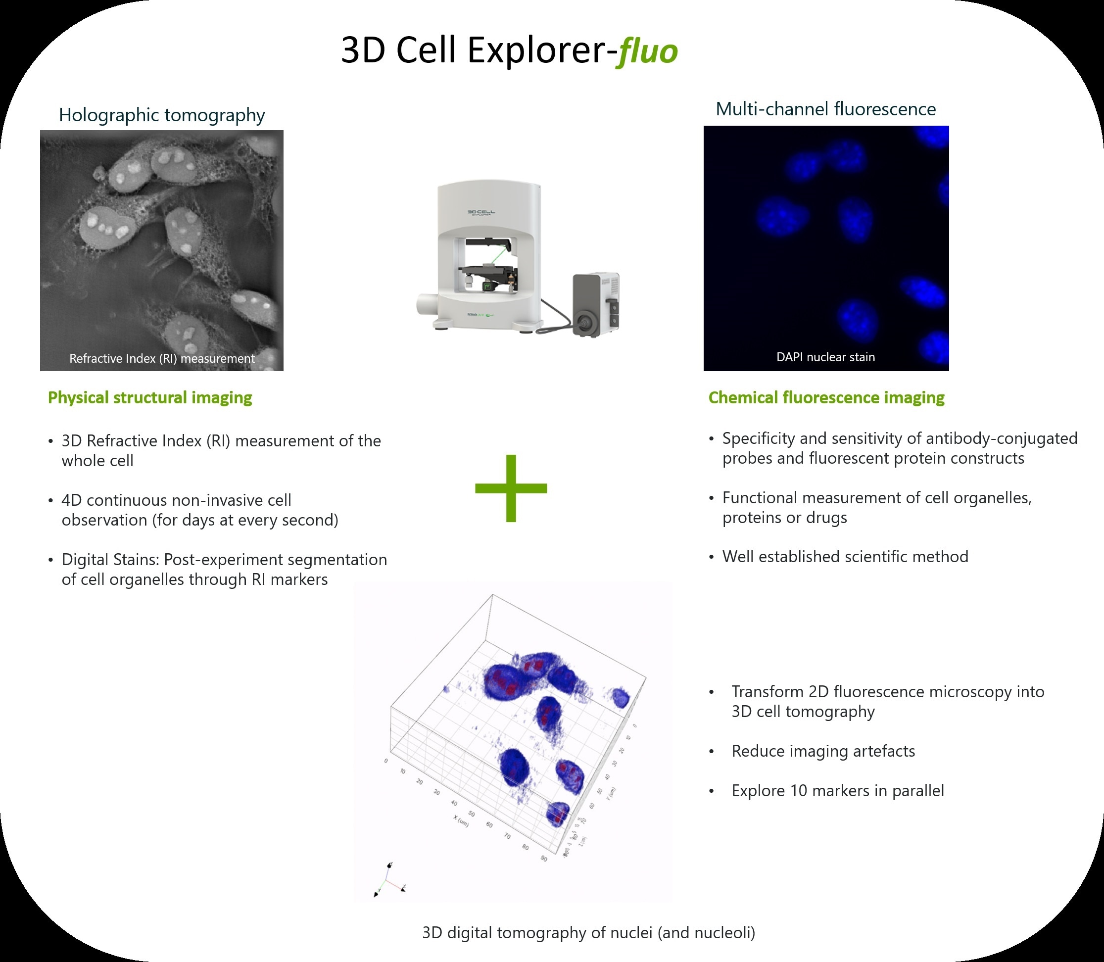
Nanolive’s newest microscope, the 3D Cell Explorer-fluo answers to the scientific need to combine the benefits from non-invasive cell tomography with a well-established and recognized method: multi-channel fluorescence microscopy. This opens completely new frontiers for research. Fluorescence microscopy offers the advantage to selectively visualize specific cell compartments by staining them with various fluorescent probes, while novel holographic tomography gives access to the whole 3D RI distribution within the cell. This combination allows to measure, for the first time, specific cell compartments in a totally non-invasive fashion, for an unlimited amount of time and in 3D. In addition, parallel time-lapse measurements of live cells with the two technologies can be used to investigate the 4D intracellular dynamics of individual organelles. It is therefore an unrivalled solution that opens the door to new dimensions for biological investigations. Researchers can now have access to two powerful imaging modalities at the same time; they can compare and correlate resulting data and add new information to their experiments.
DESIGNED AS ADVANCED RESEARCH PLATFORM: EXPLORE UP TO 10 MARKERS IN PARALLEL AT EVERY SECOND!
A key element to these unprecedented results is digital stain: the physical RI of the different cell organelles provides an inherent contrast within the cell. The overlay of the RI image of the cell with the corresponding organelle-specific fluorescence signal allows biologists to measure with high precision the exact RI of each organelle of interest and to define specifically calibrated digital stains. This process reduces the need of fluorescence markers by replacing them with up to 7 digital, non-invasive ones. Biologists are, for the first time, able to follow at the same time 7 digital markers in addition to 3 chemical ones for a total of 10 cell compartments monitored in parallel.
LIFE is in 3D! TRANSFORM 2D FLUORESCENCE MICROSCOPY INTO 3D CELL TOMOGRAPHY
Life is 3D or better 4D. The 3D Cell Explorer-fluo unveils new dimensions in cell imaging by transforming a simple 2D fluorescent signal into a complete 3D cell tomography, at every second. Since the 2D fluorescent signal allows for identification of specific organelles, the corresponding RI properties can be identified and consequently used to segment their 3D distribution in the entire cell or even multiple cells. Thus, it allows highlighting cell organelles which otherwise would be hidden by other cell compartments.
ANALYZE CELLS AS THEY ARE: ALIVE AND IN 3D
The potential impact of this technology for live cell research is enormous.
The 3D Cell Explorer-fluo produces results otherwise inaccessible. Some type of cells or cell organelles cannot be labeled because too delicate or very sensitive to bleaching or photo-toxicity. Up to now, these limitations hugely restrained cell analysis and quantification. Nanolive’s technology solves these problems by giving access to quantitative values of the whole cell. As a result, the 3D Cell Explorer and its fluorescence version bring a true revolution for live cell analysis. They allow scientists to monitor the effects of drugs and stimuli on virtually any kind of cell and without interfering with the normal cell physiology.
The 3D Cell Explorer-fluo and its older brother, the 3D Cell Explorer, really are the most powerful solutions for researchers offering groundbreaking insights into living cells, in motion and in 3D.
The 3D Cell Explorer-fluo is available now on the Nanolive webpage: https://nanolive.ch/3dcell-explorer-fluo/.
