ZEISS Lightsheet 7 Allows Multiview Imaging of Both Living and Cleared Specimens
New Light Sheet Fluorescence Microscope Introduced
Jena/Germany | ZEISS Research Microscopy Solutions
Light sheet fluorescence microscopy (LSFM) with its unique illumination principle allows fast and gentle imaging of whole living model organisms, tissues, and developing cells. The high stability of ZEISS Lightsheet 7 enables researchers to observe living samples over extended periods of time – even days – with less phototoxicity than ever before. The new light sheet microscope can also be used to image very large optically cleared specimens in toto, and with subcellular resolution. The dedicated optics, sample chambers, and sample holders of ZEISS Lightsheet 7 can be adjusted to the refractive index of the chosen clearing method to observe large samples, even whole mouse brains.
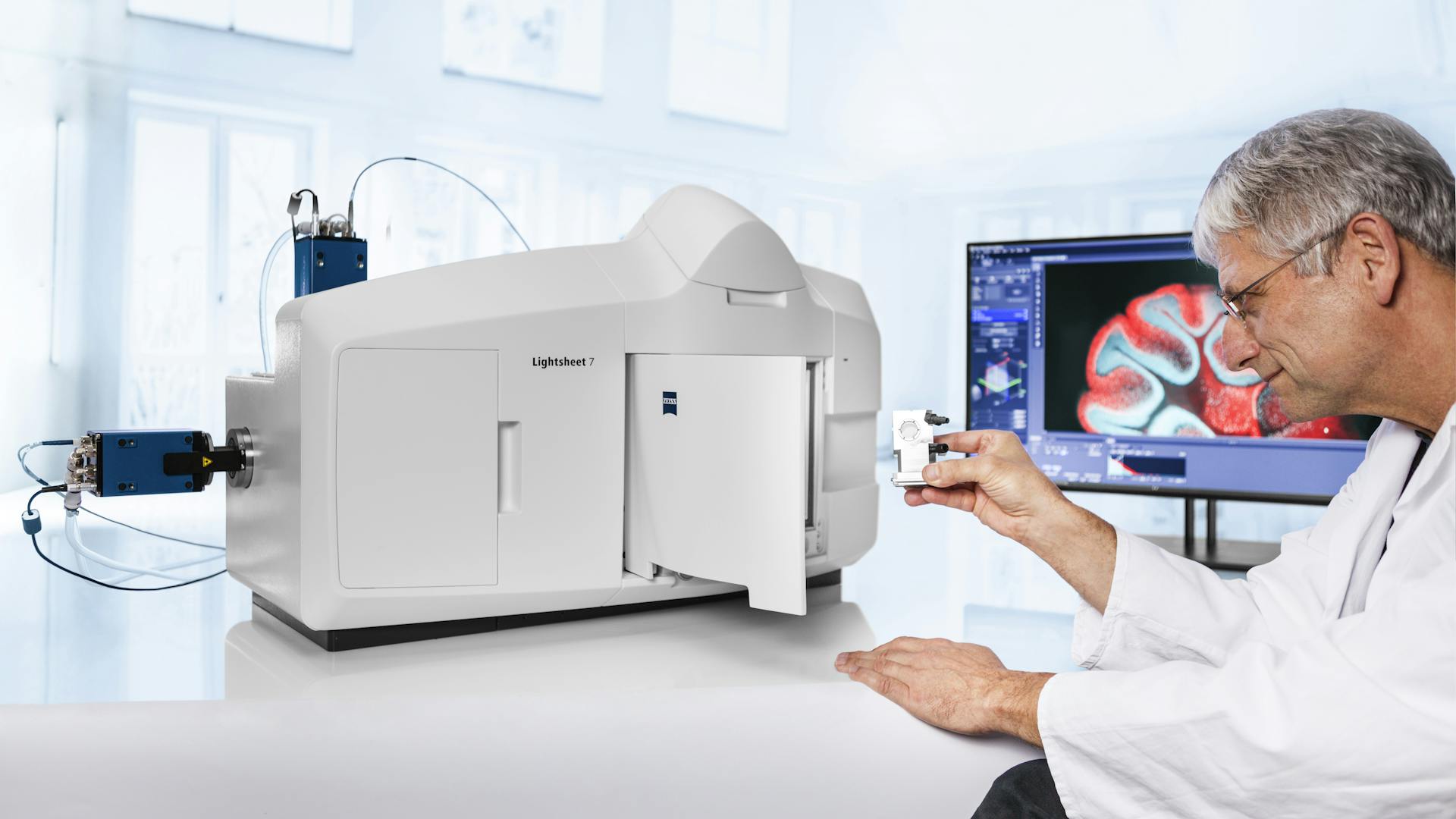
Researchers can use the exceptionally stable ZEISS Lightsheet 7 to observe living samples over extended periods of time, or to image large optically cleared specimens.
Imaging Optically Cleared Specimens
Choosing an optical clearing method depends on the type of tissue, fluorescent labels, and the size of the sample itself. ZEISS Lightsheet 7 is designed to match a multitude of conditions. Specimens with a size up to 2 cm, with a refractive index between 1.33 and 1.58, and in almost any clearing solution can be accommodated. The stable turnkey system allows acquisition of overview images as well as of data with subcellular resolution. Researchers can now perform fast and gentle LSFM imaging of optically cleared organoids, spheroids, organs, brains, or other specimens.
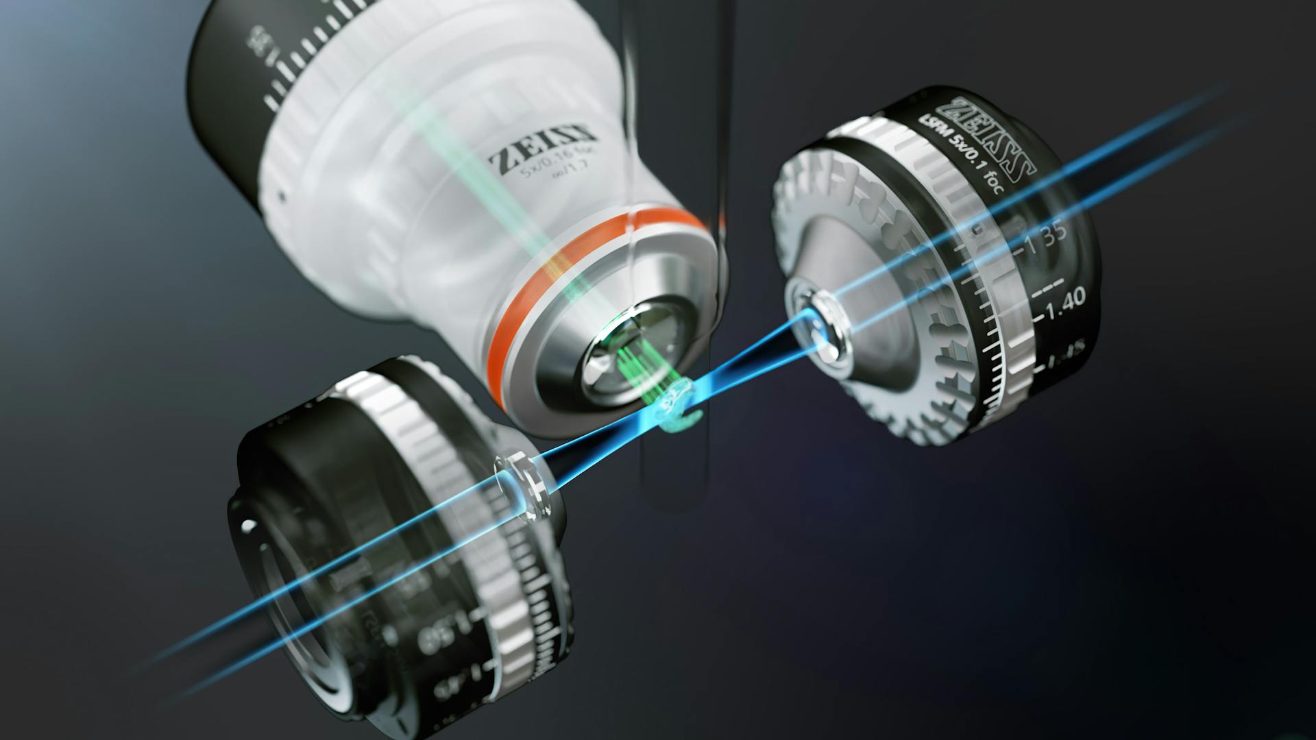
Illumination principle of ZEISS Lightsheet 7
Observing Live Samples – Fast and Gently
ZEISS Lightsheet 7 now features the high-quantum efficiency of pco.edge sCMOS detectors to enable observations of the fastest processes at the lowest illumination light levels. Scientists are able to obtain a real-life view of large samples without the adverse effects of excitation light on their biology. For vertically oriented specimens and highest frame rates, the CMOS detector ZEISS Axiocam 702 can be used. A special sample chamber provides heating, cooling, and CO2 to maintain the optimal environment for live-cell experiments. Adding Multiview and triggering options to control external devices makes it possible to observe live processes in an almost unlimited range of organisms.
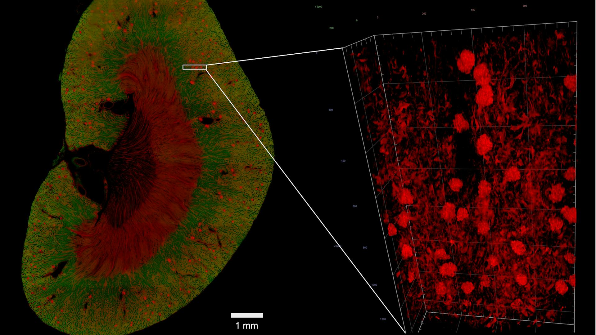
Image of a mouse kidney with ZEISS Lightsheet 7 detection optics 5× / 0.16foc. 3D whole organ imaging and computational image analysis help to gain a better understanding of the mechanisms of various kidney diseases, e.g. diabetic nephropathy. © Sample courtesy of U. Roostalu, Denmark
More Flexibility for Broader Application Range
ZEISS Lightsheet 7 brings LSFM imaging a step further to tackle a broad range of applications with best image quality. Newly designed optics and sample chambers allow users to adjust the optics to the refractive index of their samples. The new sample holder makes mounting larger specimens easier. Smart software tools assist in defining imaging parameters, such as light sheet and sample positions, the right zoom settings, tiles and positions as well as data processing parameters. All these new features go hand in hand with the reliable ZEISS combination of cylindrical lens optics and laser scanning to generate the illumination light sheet. The patented Pivot Scan technology delivers artifact-free optical sections for best possible image quality.
About ZEISS
